Date Posted, by DCP Staff
Lung cancers, the vast majority of which are caused by cigarette smoking, are the leading cause of cancer-related deaths in the United States. Lung cancer kills more people than cancers of the breast, prostate, and colon combined. By the time lung cancer is diagnosed, the disease has often already spread outside the lung. Therefore, researchers have sought to develop methods to screen for lung cancer in high-risk populations before symptoms appear. They are evaluating whether the integration of artificial intelligence – the use of computer programs or algorithms that use data to make decisions or predictions – could improve the accuracy and speed of diagnosis, aid clinical decision-making, and lead to better health outcomes.
Helical computed tomography (CT), also called low-dose CT, a technology introduced in the 1990s, can detect tumors well under 1 cm, or 0.4 inches in size, whereas chest X-rays detect tumors about 1 to 2 cm (0.4 to 0.8 inches) in size. Some researchers have hypothesized that the smaller the tumor, the higher the chance of long-term survival.
The NCI-supported National Lung Screening Trial (NLST) demonstrated that routine, annual screening of heavy smokers with low-dose CT lowered the chance of dying from lung cancer. However, there is a tradeoff: the screening increases the chance of having a false-positive screening result, which requires follow-up with a lung biopsy to rule out cancer.
The NLST, launched in 2002, compared annual screening with low-dose CT and standard chest X-ray for reducing lung cancer death rates in a high-risk population of 53,454 current and former heavy smokers. Participants were randomly assigned to undergo three annual screenings with either low-dose CT (26,722 participants) or chest X-ray (26,732 participants). The study found a 20% decrease in deaths among participants in the low-dose CT group, compared with those in the standard chest X-ray group. However, across the three annual screenings, the rate of false-positives in the low-dose CT arm was high, on average about 23%.
“Although low-dose CT screening has been shown to reduce lung cancer deaths, a high false-positive rate is a barrier to wider adoption of [this kind of] screening,” said Paul Pinsky, Ph.D., chief of NCI’s Early Detection Research Branch. “Currently, only 8-10% of those eligible are undergoing low-dose CT screening. The false-positive rate is around 20% for baseline low-dose CT screening and closer to 10% for repeat screening. This high false-positive rate results in anxiety, and follow-up imaging and can lead to invasive diagnostic work-up.”

Chief, Early Detection Research Group
NCI Division of Cancer Prevention
Artificial intelligence (AI) has been used to discriminate between normal and precancer/cancer in a number of settings. In the past decade, significant progress has been made in computer-aided detection and diagnosis, leading to several Food and Drug Administration (FDA)-approved types of software. Recent efforts are focused on deep learning, a subset of AI that uses artificial neural networks to learn from huge amounts of data. If the trained neural network out-performs human expert interpretation, it is poised to transform medical imaging and diagnostics. With the goal of reducing the high false-positive rate of low-dose CT, NCI is supporting the development of AI tools that can help radiologists better interpret these images.
“We anticipate that AI can substantially reduce the false-positive rate of low-dose CT screening while minimally affecting test sensitivity, thereby reducing diagnostic uncertainty,” Dr. Pinsky said. “But for AI research to be an effective tool for lung cancer screening, we need current high-quality image databases.”
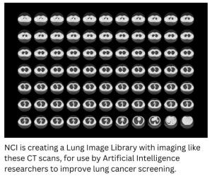
To address this need, NCI awarded a 5-year contract to Booz Allen Hamilton for the collection of medical images and data related to cancer screening. Participating medical facilities across the U.S. will collect and store low-dose CT screening and diagnostic chest CT images related to lung cancer screening and diagnostic follow-up. These images will be linked to up-to-date demographic and clinical data to serve as test and validation sets that can be shared with the research community and used by FDA.
Since the image library will not include biospecimens such as urine and blood, Dr. Pinsky and colleagues are exploring a collaboration with the new Connect for Cancer Prevention Study, a prospective cohort of 200,000 adults in nine integrative health care systems. Collection of biospecimens is needed to assess the value of adding biomarkers along with imaging for early detection of lung cancer.
“Connect would be an ideal setting where CT-scans, biospecimens, survey data, and data from electronic medical records could be integrated to evaluate low-dose CT and biomarkers.” Dr. Pinsky said. “The combination of biomarkers and AI tools may provide better predictive accuracy than either one alone.”
If you would like to reproduce some or all of this content, see Reuse of NCI Information for guidance about copyright and permissions. Please credit the National Cancer Institute as the source and link directly to the blog post using the original title, for example: "Leveraging Artificial Intelligence to Improve Accuracy of Lung Cancer Screening was originally published by the National Cancer Institute." For questions, contact us at CancerPreventionBlog@mail.nih.gov.