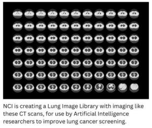
Lung cancer – the leading cause of cancer-related deaths in the United States – kills more people than cancers of the breast, prostate, and colon combined. By the time lung cancer is diagnosed, the disease has often already spread outside the lung. Researchers have sought to develop better methods to screen for lung cancer in high-risk populations before symptoms appear, but as with many technologies, refinements are needed.
Low-dose computerized tomography (CT) screening has been shown to reduce lung cancer mortality. The NCI-supported National Lung Screening Trial (NLST) showed that routine, annual screening of heavy smokers with low-dose CT lowered the chance of dying from lung cancer. However, the screening resulted in a large number of false-positive results , which required follow-up with additional imaging procedures, and sometimes invasive procedures such as lung biopsies to rule out cancer.
Leveraging Artificial Intelligence (AI) to Increase Accuracy of Lung Cancer Screening
It is anticipated that artificial intelligence (AI) – the use of computer programs, or algorithms, that use data to make decisions or predictions – can substantially reduce the false positive rate of low-dose CT screening while minimally affecting test sensitivity, thereby reducing diagnostic uncertainty. For AI research to be an effective tool for lung cancer screening, high-quality image databases are needed.
Creating a Lung Cancer Screening Image Library
To address this gap, NCI is supporting a project to create a library of low-dose CT images and corresponding data related to screening for lung cancer. Overall coordination is being supported by Booz Allen Hamilton under a larger umbrella contract with NCI for collection of medical images and data related to cancer screening.
To create this library, NCI identified several institutions across the United States that will collect low-dose CT screening and diagnostic follow-up chest CT images, along with associated clinical and demographic data. These institutions will provide geographic variation and demographic variety, along with a range of CT manufacturers.
The images will be freely available to the scientific community starting around the end of 2025.
